
استقبل المستشفى البيطري التعليمي بكلية الطب البيطري – جامعة قنا القطة «فيروز»، البالغة من العمر نحو 10 سنوات، والتي حضرت بشكوى رئيسية من زيادة ملحوظة في حجم البطن خلال الأسبوعين السابقين، مصحوبة بالخمول، وزيادة شرب المياه، واضطرابات هضمية مع وجود آثار من الدم في البراز، مع احتفاظها بشهية جيدة.
أظهر الفحص الإكلينيكي درجة حرارة في الحدود الطبيعية، مع شحوب نسبي للأغشية المخاطية وضعف النبض، بالإضافة إلى صوت غير طبيعي بالقلب (Heart Murmur)من الدرجة الرابعة من ست درجات (Grade IV/VI) مصحوبة بـ palpable precordial thrill، إلى جانب ملاحظة صعوبة في التنفس ( Dyspnea) أثناء الفحص.
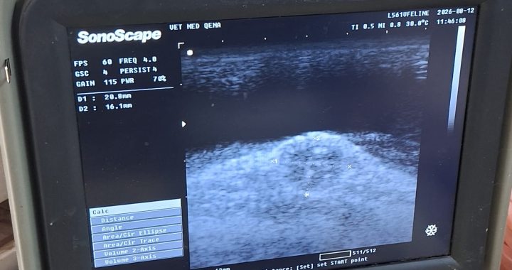
وبناءً على هذه النتائج، تم إجراء فحص شامل بالموجات فوق الصوتية للبطن، والذي أظهر كمية ملحوظة من السوائل الحرة داخل التجويف البطني (Abdominal Effusion/Ascites)، مع تقييم الكبد والمرارة والجهاز الوريدي الكبدي، بما في ذلك Hepatic Veins والـCaudal Vena Cava، كجزء من البحث عن السبب الأساسي للارتشاح.
وبناءً على نتائج الفحص الإكلينيكي والتصوير بالموجات فوق الصوتية، تم وضع Differential Diagnostic Approach يشمل الأسباب القلبية والكبدية والجهازية المحتملة، مع بدء خطة علاجية أولية داعمة شملت العلاج المدر للبول، والدعم الكبدي والجهاز الهضمي، مع تقييم إمكانية إضافة العلاج الخافض لضغط الدم وفقًا لنتائج الفحوصات وقياس ضغط الدم.
وللوصول إلى السبب الأساسي وتوجيه العلاج بصورة أدق، تم طلب مجموعة من الفحوصات المعملية شملت:
CBC – Serum Creatinine – BUN – SDMA – ALT – CPK
كما تم التوصية باستكمال التقييم القلبي باستخدام أشعة الايكو ورسم القلب Echocardiography &Electrocardiography (ECG) استكمال تقييم وتحليل السائل البطني عند الحاجة.
🔬 تعكس هذه الحالة أهمية الـClinical Reasoning والربط بين الفحص الإكلينيكي، والموجات فوق الصوتية، والتقييم القلبي، والفحوصات المعملية للوصول إلى التشخيص النهائي وتحديد الخطة العلاجية المناسبة.
مدير المستشفى
أ.د. الحسين البدري
فريق العمل :
ط.ب: عمر أحمد محمد
معيد أمراض الباطنة بقسم طب الحيوان كلية الطب البيطري جامعة قنا.
أ/ ممدوح محمد
أ/ مبارك
A Remarkable Clinical Case from the Veterinary Teaching Hospital – Qena University 🐾
The Veterinary Teaching Hospital, Faculty of Veterinary Medicine – Qena University, received “Fairoz,” a 10-year-old geriatric female cat,
presented with a chief complaint of marked abdominal enlargement over the previous two weeks, accompanied by lethargy, increased water intake, and gastrointestinal disturbances with traces of blood in the feces, while maintaining a good appetite.
Clinical examination revealed a normal body temperature, relatively pale mucous membranes and weak pulse, together with a prominent Grade IV/VI cardiac murmur accompanied by a palpable precordial thrill, in addition to noticeable respiratory abnormalities during examination.
Based on these findings, a comprehensive abdominal ultrasonographic examination was performed, revealing a marked amount of free abdominal fluid (Abdominal Effusion/Ascites). The liver, gallbladder, and hepatic vascular system were systematically evaluated, including the hepatic veins and caudal vena cava, as part of the investigation of the underlying cause of the effusion.
Based on the clinical and ultrasonographic findings, a differential diagnostic approach was established, including potential cardiovascular, hepatic, and systemic causes. An initial supportive therapeutic plan was initiated, including diuretic therapy and hepatic and gastrointestinal supportive management, while antihypertensive therapy (Amlodipine) was considered pending blood pressure measurement and laboratory results.
To further investigate the underlying cause and guide definitive management, laboratory investigations were requested, including:
CBC – Serum Creatinine – BUN – SDMA – ALT – CPK
Further cardiovascular evaluation using echocardiography and electrocardiography (ECG) was also recommended, together with further characterization and analysis of the abdominal effusion when indicated.
🔬 This case highlights the importance of clinical reasoning and integrating clinical examination, ultrasonography, cardiovascular assessment, and laboratory investigations to establish the underlying diagnosis and formulate an appropriate therapeutic plan.
Veterinary Teaching Hospital
Faculty of Veterinary Medicine – Qena University.